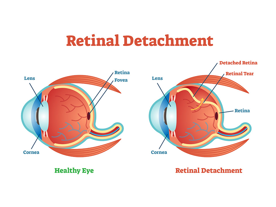
Retinal Detachment
A detached retina, or retinal detachment, is a severe eye condition that negatively impacts your visual functions and can even result in blindness if not attended to in due time.
Retinal detachment occurs when that layer of tissue behind the eyes pulls away from the tissues that support it. Some signs of this condition include seeing a shadow or floaters (squiggles and dark spots) in your vision or flashes of light.
Retinal detachment usually occurs spontaneously, and some risk factors include nearsightedness, age, history of eye trauma or surgeries, etc. If you think you have retinal detachment, visit an emergency room or contact an eye doctor immediately.
But if you are unsure, the following information should prove helpful. It will highlight what causes this condition and how to treat it. However, before we get there, let us look at what retinas do.
What Does Your Retina Do?
Your retina sends signals to your brain so you can see and sense light. However, it can’t do these jobs once it detaches. When your retina detaches, you will most likely experience some blurry vision. If it goes without treatment, you might even lose your vision permanently. So, promptly treating this condition can save your eyesight.
Causes of Retinal Detachment
The three main causes of a detached retina include;
Rhegmatogenous
The most commonly recognized cause of a detached retina occurs when there is a tiny tear in the retina. Vitreous, an eye fluid, can pass through the tear and gather behind your retina when this happens. It will then push it away, detaching the retina from the back of the eye.
This kind of detachment normally happens as one grows older. As the vitreous fluid thins and shrinks with age, it pulls on your retina, eventually tearing it.
Exudative
This is when fluid collects behind your retina, even without a retinal tear. As this fluid builds up, it pushes the retina away. The fluid buildup primarily causes swelling behind the eye. It is also caused by leaking blood vessels, which can happen because of uveitis (eye inflammation).
Tractional
This occurs when scar tissue pulls the retina away from behind the eye. Diabetes is often what causes this condition. Prolonged high blood sugar levels can damage your eye’s blood vessels, leading to scar tissue formation. The areas of traction and scars start to get bigger, detaching and pulling your retina from behind the eye.
Symptoms of Retinal Detachment
Many people do not notice any signs of a detached retina, while others do. It depends on how serious the problem is. If a huge part of your retina detaches, you will likely experience the signs and symptoms.
These symptoms can happen suddenly, and they usually include;
- Seeing many floaters, squiggly lines, dark spots, and drift flecks affects your vision. (However, seeing a few from time to time is normal and shouldn’t be a cause for concern)
- Flashes of light
- Darkened peripheral vision
- Part of your vision is covered by a shadow.
Diagnosis
To diagnose retinal detachment, you will need to undergo an eye exam. Your eye doctor will check the retina via a dilated eye exam. They will pour eye drops and then let the drops widen, or dilate, the pupil. After a couple of minutes, the doctor can look closely at your retina.
Some of the other tests your doctor might recommend after this eye exam include:
- Ocular (eye) ultrasound – This scan does not require you to take dilating drops. However, your doctor may still use them to numb the eyes, so you do not feel discomfort during the procedure. Your doctor will scan your eyes by placing a particular instrument in front of them.
- OCT (Optical coherence tomography) – This test uses dilating eye drops for imaging. Your doctor will place an OCT in front of you, and the machine will scan your eyes without touching them.
Treatment
Once you receive your diagnosis, your doctor will present you with treatment options. Your doctor may recommend a combination of two or more treatments to get the best results.
The treatments include;
Thermal (Laser) Therapy or Freezing (Cryopexy)
Sometimes, the eye doctor will spot the retinal detachment before it starts pulling away. The doctor will then use a freezing tool or medical laser to seal the tear before it happens. These medical tools create a scar that’ll hold your retina in place.
Scleral buckle
The doctor will surgically place a buckle (silicone band) around your eye during this medical procedure. The band is designed to hold your retina in place and will be there permanently. However, you will not be able to see the band. Your retinal detachment will start healing, and the doctor will finally seal the tear using a cryopexy or laser.
Pneumatic retinopexy
If your retinal detachment is not as extensive, your doctor will recommend this procedure. In this approach, the doctor will inject a tiny gas bubble into your vitreous or eye fluid. The bubble will then close the tear by pressing against your retina. You might need cryopexy or laser treatment to properly seal the tear.
Your body reabsorbs the vitreous that collects under your retina, allowing it to stick behind the eyewall as it should. The gas bubble will eventually get reabsorbed as well.
After the surgery, the doctor will instruct you to keep it still for a while, often a couple of days, to encourage healing. The doctor might also advise you to avoid lying on your back. Always follow these instructions to shorten your healing days and ensure the best outcomes.
Takeaway
Retinal detachment is a severe medical condition, but it is painless. This is why many people go a long time before they even notice they are suffering from it.
If you notice signs and symptoms of retinal detachment, like darkening of vision, flashes of light, or sudden eye floaters, you need to seek medical help as soon as possible. Visit the emergency room or get in touch with an eye doctor immediately to ensure you do not lose your eyesight.