Keratoconus is an eye condition that causes vision loss by affecting the structure of the cornea. The degenerative eye condition causes the typically spherical cornea to flatten and protrude into a cone-like form. As a result, one’s eyesight is altered.
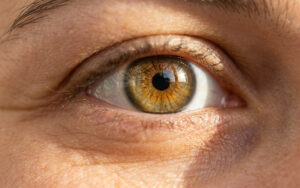
The cornea is the transparent outer layer of your eye that sits in front of your pupil and iris. The cornea is generally shaped like a ball and has a circular shape. Its primary role is to aid in the focus of light into the lens and pupil. Likewise, it also helps to keep bacteria and dirt out of your eye.
However, the cornea’s structure is not always strong enough to keep its round shape. So as light enters your eye and travels to the retina, it is deflected by the cone-shaped cornea in individuals with keratoconus.
This condition can also cause corneal edema and scarring. In addition, vision disorders such as nearsightedness (myopia) and astigmatism can also result from these alterations to your cornea.
What Causes Keratoconus?
The underlying factors that cause keratoconus are not entirely known. However, studies have shown that both hereditary and environmental factors might contribute to the development of this eye disorder.
Here are some of the factors that are thought to cause keratoconus.
1. Genetics And Family History
Genetics and family history may have a role in developing a bulging cornea. For example, some people with keratoconus have genes predisposing them to develop the condition if exposed to specific environmental factors. If you have the condition, it is always a good idea to have your children’s eyes examined.
2. Age
Keratoconus generally begins in adolescence or early adulthood. However, the condition might also develop in your late twenties or early thirties in certain circumstances.
3. Underlying Disorders
Another factor that is believed to increase the likelihood of developing keratoconus is underlying disorders. According to research, keratoconus is linked to systemic diseases such as Down syndrome, Leber congenital amaurosis, Ehlers-Danlos syndrome, brittle cornea syndrome, osteogenesis imperfecta, asthma, and retinitis pigmentosa.
4. Environmental Risk Factors
Excessive eye rubbing and improperly fitting contact lenses are environmental risk factors that may contribute to the development of keratoconus.
What are the Symptoms of Keratoconus?
The first symptom of keratoconus is an irregularly formed cornea. It seems to ‘bulge’ from the eye socket and is called astigmatism.
However, it is normal to experience no symptoms in the early stages of the disease. But as the illness worsens, your vision will become slightly distorted and blurry.
Some of the common symptoms of keratoconus that doctors look for during their eye exam include:
- Corneal swelling
- Eyestrain
- Nearsightedness
- Halos in your vision
- Light sensitivity and streaks
- Eye irritation
- Poor night vision
Are you concerned that you may be experiencing any of these symptoms? Do not hesitate to get in touch with our ophthalmologists at the Anaheim Eye Institute.
How Is Keratoconus Diagnosed?
Your eye doctor will perform a comprehensive examination, including an eye checkup and a medical and family history examination.
During your eye exam, your doctor may use the following tests to examine keratoconus:
- Corneal topography exam. This examination is done to map the curvature of your cornea. This is one of the most accurate ways to diagnose and follow up on the progression of keratoconus.
- Slit-lamp exam. During the examination, your doctor will direct a light beam to check the cornea and detect any abnormalities on the eye’s surface.
- Pachymetry. Your doctor examines the thickness of your cornea. The examination determines if the cornea is experiencing any swelling or inflammation.
What Is the Treatment for Keratoconus?
There are various options for the treatment of keratoconus. However, treatment options focus on correction of vision and depend on the condition’s stage.
1. Early Stages
Prescription Contacts or Glasses
Glasses and contact lenses are typically effective in treating myopia and astigmatism in the early stages of keratoconus. However, as keratoconus advances, glasses will no longer provide good vision, and patients may need to update their prescription lenses regularly.
2. Intermediate Stages
Custom Contact Lenses
Soft and hard contact lenses have become a common treatment for keratoconus. These custom contact lenses are used to maintain the regular shape of the cornea or reshape it to help minimize any vision distortion.
Corneal Collagen Cross-Linking Therapy
Your doctor applies a riboflavin solution to your eye. The solution increases the formation of new collagen bonds in your eye, leading to cornea recovery, preserving its shape.
Though the corneal collagen cross-link is an effective treatment for keratoconus, in some cases, it might only delay the condition’s progression. The procedure may also require the thinning of the outer layer of your cornea to improve the penetration of the riboflavin into the cornea tissue.
Are you worried about the procedure? Our team of ophthalmologists is ready to answer any questions you would have about the treatment.
3. Advanced Stages
Intracorneal Rings
Intracorneal ring segments are two crescent-shaped rings that are inserted into your cornea during surgery. The rings come in multiple varieties that a doctor prescribes for effectively reshaping your cornea. The rings are used to flatten the cornea’s surface using implantable, plastic, c-shaped ring contacts.
The surgery takes about 15-30 minutes, and you’ll be on your way in no time.
Some people may experience discomfort after the placement of the intracorneal rings. In addition, most patients still require contact lenses or glasses to correct their vision after surgery.
Corneal Transplant or Keratoplasty
The surgical procedure involves replacing the cornea after severe scarring or thinning. Cornea tissue is transplanted from a donor to replace the damaged cornea.
The procedure is performed on an outpatient basis and takes about an hour. After the procedure, you may continue to experience blurred vision, but that goes away after three to six months.
In addition, you will be required to take medication to prevent transplant rejection after the surgery. Your doctor may also prescribe contact lenses or glasses for better vision.
What’s Your Next Move?
Experiencing any eye problems? Get in touch with Anaheim Eye Institute and schedule your appointment with our team today.